An illustrated, interactive guide · Echocardiography

The Heart by Ultrasound.

Ultrasound physics, the standard views, cardiac physiology, hemodynamics, ventricular function and cardiac disease

A transducer sends a short pulse into the chest. The pulse crosses the chest wall, the right ventricle, the septum, the left ventricle and the posterior wall. Echoes return from each boundary. An amplitude (A-mode) trace and a single brightness (B-mode) line display the echoes by depth.
A sector image of the heart is acquired one scan line at a time. A selected artifact is added to the image.
An ultrasound beam intersects flowing blood at an angle. The Doppler display below shows the measured velocity, the Nyquist limit and aliasing.

Probe and body

Drag to orbit

Cross-section

Parasternal long axis

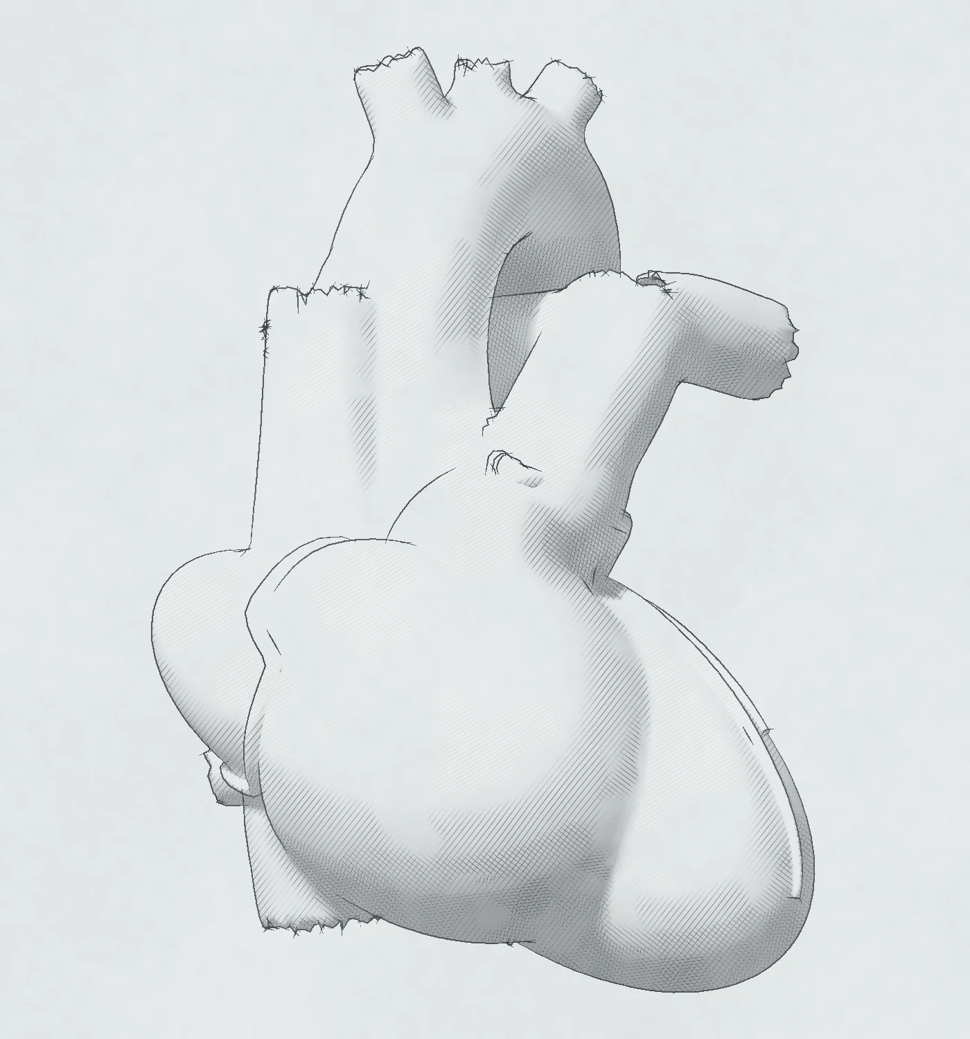
Three views of one imaging plane. Left, in the narrowest panel: the whole heart with the imaging plane and the probe, and a small figure of the body; with Probe and body off, or while the free probe is on, a three-dimensional heart with the imaging planes of the nine standard views, each in the color of its acoustic window, and the current plane highlighted. Middle: the heart cut in the current plane and seen perpendicular to it, with the structures labeled. Right: the echocardiographic image of the same plane.
Wiggers diagram of one heartbeat: aortic, left ventricular and left atrial pressure, left ventricular volume, the ECG, the heart sounds, mitral inflow and left ventricular outflow Doppler, and an apical four-chamber frame at the chosen moment.
Left ventricular pressure-volume loop with the end-systolic and end-diastolic pressure-volume relations and the arterial elastance line, beside the normal loop.
Two settings. Flow and pressure, at the left: blood flows from the LV outflow tract through the aortic valve into the aorta. The same flow passes every level, so the velocity rises where the area is small, and the pressure falls there and partly rises again in the aorta. Strips under the tube show the flow, the velocity and the pressure along it, and pulsed-wave Doppler before the valve and continuous-wave Doppler through it share one velocity scale. IVC, at the left: the subcostal long axis shows the IVC through the liver into the right atrium, with a caliper 1 to 2 cm from the right atrium, and an M-mode recording shows the IVC through quiet breathing and a sniff, with its largest and smallest diameters. At the right in both settings: right ventricular systolic pressure from the tricuspid regurgitation velocity, as a bar that stacks the pressure difference on the right atrial pressure that the IVC gives.
Apical four-chamber and two-chamber echo views with the left ventricular border traced, its long axis and twenty disks for the biplane method, each above a drawing of the same ventricle divided into the same disks; or, for the right ventricle, TAPSE on M-mode, tricuspid annular velocity and fractional area change.
Occluded artery
Buttons for a normal heart and for an occluded LAD, RCA or circumflex artery. Seventeen-segment bullseye colored by the wall motion score of each segment, with the occluded artery drawn on it, and the apical four-, two- and three-chamber echo views, in which each segment moves according to its score. The numbered segment buttons under the controls change the scores.
Diastolic function from normal to restrictive filling: an apical four-chamber view with the left atrium to scale, mitral inflow, septal and lateral tissue Doppler, pulmonary vein flow, left atrial strain and the tricuspid jet, with the 2025 diastolic algorithm and the path that the current values follow through it.
Cardiac masses from none to large: a left atrial myxoma on a stalk from the atrial septum, which prolapses through the mitral valve in diastole, in the apical four-chamber view and the parasternal long axis, with continuous-wave mitral inflow and its mean gradient; an LV thrombus on an akinetic apex, mural, protruding or mobile, in the apical four-chamber view and the short axis at the apex, with the wall motion of the 17 segments; a papillary fibroelastoma on the aortic side of an aortic cusp, in the parasternal and apical long axes zoomed on the valve. Calipers measure each mass. With an ultrasound enhancing agent the cavities fill, and each mass shows its uptake.
A continuous-flow left ventricular assist device at pump speeds from 4,600 to 6,200 rpm, with a working pump, right ventricular failure, de novo aortic regurgitation or an obstruction of the outflow graft: a parasternal long-axis view with calipers on the left ventricle and color jets of aortic and mitral regurgitation, and an apical four-chamber view with the inflow cannula in the left ventricular apex, the flow into it and the mitral and tricuspid jets, both cut from the heart model; pulsed-wave Doppler at the inflow cannula and continuous-wave Doppler in the outflow graft over three beats, with the artificial pulse every 2 seconds; pulsed-wave Doppler in the right ventricular outflow tract with its velocity time integral and the cardiac output; an M-mode through the aortic cusps over the same three beats; and the left ventricular end-diastolic diameter plotted against the pump speed.
Aortic stenosis from a normal valve to severe stenosis and then low flow: an apical five-chamber view with the LV outflow tract, the thickened and calcified valve, the aortic root and the jet, and the continuous-wave Doppler line through the valve; the continuous-wave trace with the dense LVOT envelope inside the aortic jet and both peak velocities marked; the short-axis valve orifice to scale; and the measurements against their cut-offs.
Aortic regurgitation from mild to severe: a parasternal long-axis view with the color jet in the outflow tract and a dilating left ventricle, the continuous-wave jet with its pressure half-time, flow reversal in the descending aorta, outflow Doppler, and the measurements against their cut-offs.
Mitral stenosis from a normal valve to severe stenosis: an apical four-chamber view with doming leaflets, a large left atrium and the color inflow jet with its flow convergence, the short-axis orifice to scale, continuous-wave mitral inflow with the pressure half-time, the tricuspid jet, and the measurements against their cut-offs.
Mitral regurgitation from mild to severe: an apical four-chamber view with the color jet and flow convergence, the continuous-wave jet, pulmonary vein and mitral inflow Doppler, and the measurements against their cut-offs.
Tricuspid regurgitation from mild to torrential: an apical four-chamber view with the color jet and a dilating right heart, the continuous-wave jet, hepatic vein flow, the inferior vena cava on M-mode, a short-axis view of the septum, and the measurements against their cut-offs.
A prosthetic aortic valve from normal function to significant stenosis, or a normal valve that is small for the patient: a zoomed parasternal long-axis view and a short-axis view of the valve, cut from the heart model, with the sewing ring and the stent and leaflets of a bioprosthesis or the housing and discs of a bileaflet mechanical valve, and the shadow and reverberation behind the metal; continuous-wave Doppler through the valve with its peak velocity, acceleration time and ejection time; pulsed-wave Doppler in the LVOT; and the measurements against their cut-offs.
Transcatheter aortic valve replacement for aortic stenosis, step by step: the mid-esophageal aortic valve long-axis and short-axis views of transesophageal echo, cut from the heart model. Measure: the calcified native valve and the annulus from hinge point to hinge point. Cross: the guidewire and the crimped valve over the annulus. Deploy: a balloon-expandable or a self-expanding frame that expands and pushes the native leaflets out. Check: color Doppler of paravalvular jets outside the frame in diastole and their share of the circumference; continuous-wave Doppler through the valve and pulsed-wave Doppler in the LVOT from the deep transgastric view; and the measurements against the criteria of a successful result.
Diseases of the aorta, for dilation, dissection or coarctation. Dilation, from normal to 6.0 cm at the sinuses and the ascending aorta: the parasternal long-axis view zoomed on the aortic root, cut from the heart model, at end-diastole with leading-edge calipers on the sinuses of Valsalva, the sinotubular junction and the ascending aorta, and at midsystole with an inner-edge caliper on the annulus; the four diameters against their normal ranges; and the diameter thresholds of the 2022 ACC/AHA guideline, for a bicuspid valve under the bicuspid heart. Dissection, from no flap through a reverberation artifact to a flap with aortic regurgitation and a pericardial effusion: the parasternal long-axis view with color, the flap between the true and false lumens, brisk forward flow in the true lumen and slow, late, reversed flow in the false lumen, and a regurgitant jet whose origin and direction follow the flap: central as the root dilates, eccentric from a prolapsed cusp, and beside the flap when it prolapses through the valve; an M-mode of the ascending aorta; and the root zoomed, from one interspace higher. Coarctation, from a mild narrowing to severe coarctation: the suprasternal view of the arch with the narrowing just past the left subclavian artery, its caliper, the color jet and the continuous-wave beam; the subcostal view of the abdominal aorta with its diameter and the pulsed-wave gate; continuous-wave Doppler across the narrowing with the diastolic tail; pulsed-wave Doppler of the abdominal aorta; and the measurements against their cut-offs.
Pericardial disease, with a switch for the disease. Pericardial effusion from none to tamponade: an apical four-chamber view with the fluid around the heart and, in tamponade, collapse of the right atrium and right ventricle; mitral inflow, tricuspid inflow and hepatic vein flow through one respiratory cycle; a parasternal long-axis view with the fluid behind the left ventricle and, in a large effusion, in front of the right ventricle; the inferior vena cava on M-mode; and the measurements against their cut-offs. Constrictive pericarditis from a normal pericardium to severe constriction: an apical four-chamber view with the pericardium at its measured thickness and the septum shifting with each breath; mitral and tricuspid inflow and hepatic vein flow over two breaths with a respiratory trace; tissue Doppler of the medial and lateral mitral annulus; an M-mode of the septum with breathing; the inferior vena cava on M-mode; and the measurements against their cut-offs.
Cardiac amyloidosis from a normal heart to advanced disease: a parasternal long-axis view at end-diastole with calipers on the septum, the cavity and the inferolateral wall, and an apical four-chamber view, both cut from the heart model; mitral inflow and septal tissue Doppler; the 17-segment longitudinal strain bull's-eye; and the measurements against their cut-offs.
Hypertrophic cardiomyopathy from a normal heart to severe obstruction, at rest or with the Valsalva maneuver: a parasternal long-axis view with calipers on the septum, the cavity and the posterior wall at end-diastole and the gap between the anterior mitral leaflet and the septum in systole, and an apical three-chamber view with the color jets in the LV outflow tract and the left atrium, both cut from the heart model; continuous-wave Doppler of the outflow tract and of the mitral regurgitation on one velocity scale; and an M-mode through the mitral valve that shows systolic anterior motion.
Myocardial strain from normal function to severe dysfunction, in a global, a regional or an apical-sparing pattern: the apical four-, two- and three-chamber views cut from the heart model, each with the myocardial midline of speckle tracking in six segments colored by their strain; the strain curves of the chosen view through one beat, with aortic valve closure; the 18-segment bull's-eye with GLS; and the arithmetic of the chosen view's strain, with the relative apical LS in the apical-sparing pattern. Cardio-oncology: a baseline study and a follow-up study during cancer therapy side by side, each with its four-chamber view, LVEF, bull's-eye and GLS, and the result by the ESC 2022 definitions of cancer therapy-related cardiac dysfunction.
View
Buttons for the apical four-, two- and three-chamber views. Dobutamine stress echocardiography as a quad screen: the chosen view at rest, at low dose, at peak and in recovery, each cut from the heart model and beating at the heart rate of its stage, with the segment numbers on its walls colored by their wall motion score. Beside it, the response of each of the 17 segments on a bull's-eye, and the protocol: the dobutamine dose in 3-minute stages, atropine, and the heart rate against its target.
Shunts from none to large, for a secundum atrial septal defect, a perimembranous ventricular septal defect or a patent foramen ovale. Atrial septal defect: a subcostal view of the atrial septum with the defect, its caliper and the left-to-right color jet; an RV-focused apical four-chamber view with the RV and RA size; pulsed-wave Doppler in the RV and LV outflow tracts with their VTIs and diameters for Qp/Qs; and the continuous-wave TR jet with the RV systolic pressure. Ventricular septal defect: a short-axis view at the aortic valve with the defect just below the valve at about 11 o'clock and its jet into the RV; a parasternal long-axis view with the LV diameter; continuous-wave Doppler through the defect with its peak velocity, gradient and the RV systolic pressure; and the two outflow tracts for Qp/Qs. Patent foramen ovale: an apical four-chamber view during an agitated saline study, with the right heart opacified and bubbles crossing into the left atrium, and the number of bubbles in the left atrium beat by beat, at rest and after Valsalva release. LA pressure: a subcostal view of a restrictive 5-mm atrial communication with its left-to-right color jet; the LA and RA pressures over two beats, with their means; and continuous-wave Doppler across the septum, whose peak velocity and traced envelope, with the RA pressure, give the peak and mean LA pressure.
The right ventricle from normal to severe overload, under a pressure or a volume load, or in acute pulmonary embolism: an RV-focused apical four-chamber view with the RV size, wall and FAC, a short-axis view of the septum with the two diameters of the eccentricity index, the continuous-wave TR jet with the RV systolic pressure, pulsed-wave flow in the RV outflow tract with its acceleration time, the M-mode of TAPSE and the tissue Doppler S′. In acute pulmonary embolism the four-chamber view also gives the RV/LV ratio and the free wall's inward motion at its base, middle and apex, with McConnell's sign; the TR jet gives the TR gradient, and the two Doppler traces mark the 60/60 sign when it holds.
Infective endocarditis from a normal mitral valve to a large vegetation with leaflet perforation and severe regurgitation: the parasternal long-axis view zoomed on the valves, with the vegetation on the atrial side of the anterior mitral leaflet and a caliper on its maximal length; the apical three-chamber view with the color jet through the perforation; continuous-wave Doppler through the mitral valve; pulmonary vein flow; and the measurements against their cut-offs.
Mechanical complications of myocardial infarction from none to the end stage, one at a time: a rupture of the apical septum in the apical four-chamber view with its left-to-right color jet, the CW jet across it and the PW velocities of the two outflow tracts for Qp/Qs; a papillary muscle rupture with a flail anterior mitral leaflet, the torn head of the muscle and an eccentric MR jet in the apical three-chamber view, with the CW MR jet and the pulmonary vein flow; a pseudoaneurysm of the inferolateral wall in the parasternal long axis, with calipers on its neck and sac, and the pericardial effusion and the signs of tamponade in the apical four-chamber view; a true apical aneurysm in the apical four- and two-chamber views, with calipers on its mouth and width, a mural thrombus at the end stage, and the wall motion of the 17 segments.
Mitral transcatheter edge-to-edge repair of a flail posterior leaflet, step by step, from the transthoracic study before the procedure to the one after it. Left: the heart model cut away, with the guide from the inferior vena cava across the atrial septum and the clip on the mitral valve, at the step shown. Right, at each step, the images that guide it: before, the parasternal long axis and the TEE long-axis view with the flail P2 and its eccentric jet, and the 3D en face view of the valve; the transseptal puncture in the bicaval view, its short-axis view and the four-chamber view with the height of the puncture site above the leaflets; steering, alignment and grasping in the commissural and long-axis X-plane views and the 3D en face view; the assessment before release with the residual jets, the continuous-wave Doppler of the inflow with its mean gradient and the transgastric short axis of the double orifice; the release with the left-to-right flow across the iatrogenic atrial septal defect; and after the procedure, the parasternal short axis of the double orifice, the apical four-chamber view with the residual jets and the inflow's mean gradient.
Tricuspid transcatheter edge-to-edge repair (T-TEER) of massive secondary tricuspid regurgitation, step by step, from the transthoracic study before the procedure to the one after it. Left: the heart model cut away, with the guide from the inferior vena cava into the right atrium and the clip on the anteroseptal line of coaptation, at the step shown. Right, at each step, the images that guide it: before, the RV-focused apical four-chamber view and the parasternal short axis of the tricuspid valve with the central coaptation gap and the wide jet, and continuous-wave Doppler of the regurgitation; the guide in the right atrium in the four-chamber and RV inflow-outflow views with the 3D en face view of the valve from the right atrium; steering, alignment and grasping in the mid-esophageal X-plane views along and across the line of coaptation, the transgastric short axis of the valve and the 3D en face view; the assessment before release with the residual jet in the four-chamber view, the continuous-wave Doppler of the inflow with its mean gradient and the transgastric short axis of the orifices; the release; and after the procedure, the RV-focused four-chamber view with the residual jet, the hepatic vein flow and the inflow's mean gradient.
Automated measurement by EchoNet-Measurements, drawn on the site's own images. B-mode: the view of the chosen measurement, cut from the heart model, with a heat map on each end of the caliper, the caliper between their weighted centers and its length on each frame; a close-up of one heat map on its pixels with its weighted center; the conversion from pixels to centimeters; and a trace of the measurement over three beats with end-diastole and end-systole marked and the reported value circled. With the two-peaked map, one heat map has a second peak on a nearby structure and its weighted center falls between the peaks. Doppler and M-mode: the recording of the chosen measurement from its chapter, with a heat map on each peak (two for E and A, two for TAPSE), the baseline, a close-up of one map and the conversion from pixels to velocity or distance. Without the DICOM data, the points remain and the figure gives pixels only.
How EchoPrime and EchoNet-Measurements read one study of the heart shown, in seven steps. EchoPrime: the 14 videos of the study, cut from the heart model, each tagged with its view; one video encoder that turns each video into 512 numbers, drawn as a stripe beside the video's view; the public attention weights of the chosen section, a line from each video to the section as wide as its weight, with the weight at its start, under a strip of the 15 sections; and the chosen section's task heads, of about 150: each number head with its value, ringed where it checks a measurement, then the words of its word heads. EchoNet-Measurements: the nine B-mode measurements, each on its view at the frame that it reports, with its heat maps and its caliper, and the nine Doppler and M-mode measurements on eight recordings, each with its value. Then the measurements of the chosen section beside one band, the heads' range of ± 10%, with a dot for each caliper and its value. Last, the report for the heart, with the words in a serif face and the measured numbers in blue. A click on the strip of sections, a tile or a sentence chooses a section. Arrows link each step to the next. The anchor sits beside the measurements, and the report runs across the foot of the figure. On a small screen, the steps run in more rows, and the figure scrolls.

Probe and plane

Drag to orbit

ME four-chamber

Two views of one transesophageal imaging plane. Left: the three-dimensional heart seen from the patient's left, with the esophagus behind the left atrium, the probe in it at the chosen level, and the imaging plane, which turns with the multiplane angle. Right: the echocardiographic image of the same plane in the guideline's display orientation, with the structures labeled, the view's name, the image depth and an icon of the multiplane angle.

Regional wall motion

The 17-segment model

In the standard model, the LV wall is divided into 17 segments. Each coronary artery usually supplies a specific set of segments, so the distribution of abnormal wall motion indicates the artery involved.

Wall motion of the affected segments
Each segment: tap for the next score

Segment nomenclature

The segments are named counterclockwise from the anterior junction of the septum and the RV free wall: anteroseptal, inferoseptal, inferior, inferolateral, anterolateral and anterior14. The basal and mid levels each contain six segments, the apical level contains four, and the apical cap is segment 1714.

  • Wall motion should be scored in 16 segments, and 17 segments should be used for perfusion or for comparison with other imaging modalities14.
  • Each segment is scored as follows: 1, normal or hyperkinetic; 2, hypokinetic; 3, akinetic; 4, dyskinetic14. The 2025 reporting guideline adds a score of 5 for an aneurysmal segment36. In the figure, a tap on a segment in the bull's-eye or in an apical view, or on its numbered button, advances that segment to the next score.
  • The wall motion score index is the mean of the segment scores, and a value of 1.00 is normal14.

Apical views and LV walls

The apical four-chamber view shows the inferoseptal and anterolateral walls, the two-chamber view the inferior and anterior walls, and the three-chamber view the inferolateral and anteroseptal walls14.

Wall motion patterns of coronary occlusion

  • LAD. LAD occlusion affects the anterior wall in the two-chamber view and the anteroseptal wall in the three-chamber view, each from base to apex; the apical septum in the four-chamber view; and the apex in all three views4114.
  • RCA. RCA occlusion affects the inferior wall in the two-chamber view, from base to apex, and the basal and mid inferoseptum in the four-chamber view; the three-chamber view remains normal4114.
  • Circumflex. Circumflex occlusion affects the anterolateral wall in the four-chamber view and the inferolateral wall in the three-chamber view; the two-chamber view remains normal4114.

Coronary territories

In the standard map, the LAD supplies segments 1, 2, 7, 8, 13, 14 and 17; the right coronary artery supplies segments 3, 4, 9, 10 and 15; and the circumflex supplies segments 5, 6, 11, 12 and 1641. Coronary supply varies between patients; in one CT study, 72% of patients had at least one segment supplied by a different artery41. The map should therefore be regarded as a guide rather than a fixed rule.

Regional strain values are not recommended for clinical decision-making, because they vary between repeated examinations and between vendors39.

Key points

  1. Wall motion is scored in 16 segments, and the wall motion score index is the mean of their scores.
  2. Each apical view shows two opposite walls of the LV.
  3. Abnormal motion of the anterior wall, the anteroseptum and the apex suggests LAD occlusion. Abnormal motion of the inferior wall and the basal and mid inferoseptum suggests RCA occlusion, and abnormal motion of the lateral walls suggests circumflex occlusion.

By David Ouyang, MD · Updated

Chapters

    Sources

    Each guideline value on this page is cited to its source. The cut-off values are taken from the American Society of Echocardiography (ASE), the European Association of Cardiovascular Imaging (EACVI), the British Society of Echocardiography (BSE), the ACC/AHA, the ESC and the ESC/ERS.

    The figures are generated by simple models calibrated to agree with these sources. The diastolic values and the Doppler and strain values of the amyloid heart are illustrative and are not patient data. All images are simulated, including the drawings, the heart model and the echocardiographic views.

    Sources, in reading order

    1. Lacefield JC. Physics of ultrasound; Ultrasound imaging. In: Dance DR, Christofides S, Maidment ADA, et al, eds. Diagnostic Radiology Physics: A Handbook for Teachers and Students. Vienna: International Atomic Energy Agency; 2014:291-331. www-pub.iaea.org
    2. Efrimescu CI, Ng WCK, Vegas A. Perioperative 3D transoesophageal echocardiography. Part 1: fundamental principles. BJA Educ 2024;24:217-226. doi:10.1016/j.bjae.2024.03.001 · PMID 38764440
    3. Urone PP, Hinrichs R. College Physics 2e. Section 17.7: Ultrasound. Houston (TX): OpenStax; 2022. ISBN 978-1-951693-60-2 (digital); 978-1-711470-83-2 (print). openstax.org
    4. Mitchell C, Rahko PS, Blauwet LA, et al. Guidelines for Performing a Comprehensive Transthoracic Echocardiographic Examination in Adults: Recommendations from the American Society of Echocardiography. J Am Soc Echocardiogr 2019;32:1-64. doi:10.1016/j.echo.2018.06.004 · PMID 30282592
    5. Zander D, Hüske S, Hoffmann B, et al. Ultrasound Image Optimization ("Knobology"): B-Mode. Ultrasound Int Open 2020;6:E14-E24. doi:10.1055/a-1223-1134 · PMID 32885137
    6. Bertrand PB, Levine RA, Isselbacher EM, et al. Fact or Artifact in Two-Dimensional Echocardiography: Avoiding Misdiagnosis and Missed Diagnosis. J Am Soc Echocardiogr 2016;29:381-391. doi:10.1016/j.echo.2016.01.009 · PMID 26969139
    7. Starkoff B. Ultrasound physical principles in today's technology. Australas J Ultrasound Med 2014;17:4-10. doi:10.1002/j.2205-0140.2014.tb00086.x · PMID 28191202
    8. Hawkins K, Henry JS, Krasuski RA. Tissue harmonic imaging in echocardiography: better valve imaging, but at what cost? Echocardiography 2008;25:119-123. doi:10.1111/j.1540-8175.2007.00575.x · PMID 18269555
    9. Quiñones MA, Otto CM, Stoddard M, et al. Recommendations for quantification of Doppler echocardiography: a report from the Doppler Quantification Task Force of the Nomenclature and Standards Committee of the American Society of Echocardiography. J Am Soc Echocardiogr 2002;15:167-184. doi:10.1067/mje.2002.120202 · PMID 11836492
    10. Paeng DG, Lee CA, Imtiaz C. Principles of Doppler ultrasound and emerging blood flow imaging. Ultrasonography 2025;44:409-424. doi:10.14366/usg.25152 · PMID 41157819
    11. Robinson S, Rana B, Oxborough D, et al. A practical guideline for performing a comprehensive transthoracic echocardiogram in adults: the British Society of Echocardiography minimum dataset. Echo Res Pract 2020;7:G59-G93. doi:10.1530/ERP-20-0026 · PMID 33112828
    12. Mitchell JR, Wang JJ. Expanding application of the Wiggers diagram to teach cardiovascular physiology. Adv Physiol Educ 2014;38:170-175. doi:10.1152/advan.00123.2013 · PMID 24913453
    13. Alhakak AS, Olsen FJ, Skaarup KG, et al. Age- and sex-based normal reference ranges of the cardiac time intervals: the Copenhagen City Heart Study. Clin Res Cardiol 2025;114:430-442. doi:10.1007/s00392-023-02269-2 · PMID 37522900
    14. Lang RM, Badano LP, Mor-Avi V, et al. Recommendations for Cardiac Chamber Quantification by Echocardiography in Adults: An Update from the American Society of Echocardiography and the European Association of Cardiovascular Imaging. J Am Soc Echocardiogr 2015;28:1-39.e14. doi:10.1016/j.echo.2014.10.003 · PMID 25559473
    15. Voigt JU, Pedrizzetti G, Lysyansky P, et al. Definitions for a Common Standard for 2D Speckle Tracking Echocardiography: Consensus Document of the EACVI/ASE/Industry Task Force to Standardize Deformation Imaging. J Am Soc Echocardiogr 2015;28:183-193. doi:10.1016/j.echo.2014.11.003 · PMID 25623220
    16. Schimpf R, Antzelevitch C, Haghi D, et al. Electromechanical coupling in patients with the short QT syndrome: further insights into the mechanoelectrical hypothesis of the U wave. Heart Rhythm 2008;5:241-245. doi:10.1016/j.hrthm.2007.10.015 · PMID 18242547
    17. Nagueh SF, Sanborn DY, Oh JK, et al. Recommendations for the Evaluation of Left Ventricular Diastolic Function by Echocardiography and for Heart Failure With Preserved Ejection Fraction Diagnosis: An Update From the American Society of Echocardiography. J Am Soc Echocardiogr 2025;38:537-569. doi:10.1016/j.echo.2025.03.011 · PMID 40617625
    18. Bastos MB, Burkhoff D, Maly J, et al. Invasive left ventricle pressure-volume analysis: overview and practical clinical implications. Eur Heart J 2020;41:1286-1297. doi:10.1093/eurheartj/ehz552 · PMID 31435675
    19. Suga H, Sagawa K, Shoukas AA. Load independence of the instantaneous pressure-volume ratio of the canine left ventricle and effects of epinephrine and heart rate on the ratio. Circ Res 1973;32:314-322. doi:10.1161/01.res.32.3.314 · PMID 4691336
    20. Kelly RP, Ting CT, Yang TM, et al. Effective arterial elastance as index of arterial vascular load in humans. Circulation 1992;86:513-521. doi:10.1161/01.cir.86.2.513 · PMID 1638719
    21. Monge García MI, Santos A. Understanding ventriculo-arterial coupling. Ann Transl Med 2020;8:795. doi:10.21037/atm.2020.04.10 · PMID 32647720
    22. Burkhoff D, Sagawa K. Ventricular efficiency predicted by an analytical model. Am J Physiol 1986;250:R1021-R1027. doi:10.1152/ajpregu.1986.250.6.R1021 · PMID 3717375
    23. Chantler PD, Lakatta EG. Arterial-Ventricular Coupling with Aging and Disease. Front Physiol 2012;3:90. doi:10.3389/fphys.2012.00090 · PMID 22586401
    24. Antonini-Canterin F, Poli S, Vriz O, et al. The Ventricular-Arterial Coupling: From Basic Pathophysiology to Clinical Application in the Echocardiography Laboratory. J Cardiovasc Echogr 2013;23:91-95. doi:10.4103/2211-4122.127408 · PMID 28465893
    25. Neema PK, Panidapu N. The Mechanisms and Pathophysiology of Mitral Regurgitation: A Narrative Review. Ann Card Anaesth 2025;28:109-118. doi:10.4103/aca.aca_221_24 · PMID 40237656
    26. Klabunde RE. Mitral regurgitation. Cardiovascular Physiology Concepts (cvphysiology.com). Revised 02/03/2024. cvphysiology.com
    27. Vincent JL. Understanding cardiac output. Crit Care 2008;12:174. doi:10.1186/cc6975 · PMID 18771592
    28. Norton JM. Toward consistent definitions for preload and afterload. Adv Physiol Educ 2001;25:53-61. doi:10.1152/advances.2001.25.1.53 · PMID 11824209
    29. Zoghbi WA, Adams D, Bonow RO, et al. Recommendations for Noninvasive Evaluation of Native Valvular Regurgitation: A Report from the American Society of Echocardiography Developed in Collaboration with the Society for Cardiovascular Magnetic Resonance. J Am Soc Echocardiogr 2017;30:303-371. doi:10.1016/j.echo.2017.01.007 · PMID 28314623
    30. Baumgartner H, Hung J, Bermejo J, et al. Recommendations on the echocardiographic assessment of aortic valve stenosis: a focused update from the European Association of Cardiovascular Imaging and the American Society of Echocardiography. J Am Soc Echocardiogr 2017;30:372-392. doi:10.1016/j.echo.2017.02.009 · PMID 28385280
    31. Humbert M, Kovacs G, Hoeper MM, et al. 2022 ESC/ERS Guidelines for the diagnosis and treatment of pulmonary hypertension. Eur Heart J 2022;43:3618-731. doi:10.1093/eurheartj/ehac237 · PMID 36017548
    32. Mukherjee M, Rudski LG, Addetia K, et al. Guidelines for the echocardiographic assessment of the right heart in adults and special considerations in pulmonary hypertension: recommendations from the American Society of Echocardiography. J Am Soc Echocardiogr 2025;38:141-86. doi:10.1016/j.echo.2025.01.006 · PMID 40044341
    33. Rudski LG, Lai WW, Afilalo J, et al. Guidelines for the echocardiographic assessment of the right heart in adults: a report from the American Society of Echocardiography. J Am Soc Echocardiogr 2010;23:685-713. doi:10.1016/j.echo.2010.05.010 · PMID 20620859
    34. Tucker WD, Shrestha R, Burns B. Anatomy, Abdomen and Pelvis: Inferior Vena Cava. In: StatPearls. Treasure Island (FL): StatPearls Publishing; 2023 (updated July 24, 2023). PMID 29493975 · ncbi.nlm.nih.gov
    35. Assavapokee T, Rola P, Assavapokee N, Koratala A. Decoding VExUS: a practical guide for excelling in point-of-care ultrasound assessment of venous congestion. Ultrasound J 2024;16:48. doi:10.1186/s13089-024-00396-z · PMID 39560910
    36. Taub CC, Stainback RF, Abraham T, et al. Guidelines for the standardization of adult echocardiography reporting: recommendations from the American Society of Echocardiography. J Am Soc Echocardiogr 2025;38:735-74. doi:10.1016/j.echo.2025.06.001 · PMID 40912865
    37. Heidenreich PA, Bozkurt B, Aguilar D, et al. 2022 AHA/ACC/HFSA guideline for the management of heart failure: a report of the American College of Cardiology/American Heart Association Joint Committee on Clinical Practice Guidelines. Circulation 2022;145:e895-e1032. doi:10.1161/CIR.0000000000001063 · PMID 35363499
    38. Otto CM, Nishimura RA, Bonow RO, et al. 2020 ACC/AHA guideline for the management of patients with valvular heart disease: a report of the American College of Cardiology/American Heart Association Joint Committee on Clinical Practice Guidelines. Circulation 2021;143:e72-e227. doi:10.1161/CIR.0000000000000923 · PMID 33332150
    39. Thomas JD, Edvardsen T, Abraham T, et al. Clinical applications of strain echocardiography: a clinical consensus statement from the American Society of Echocardiography developed in collaboration with the European Association of Cardiovascular Imaging of the European Society of Cardiology. J Am Soc Echocardiogr 2025;38:985-1020. doi:10.1016/j.echo.2025.07.007 · PMID 40864001
    40. Thavendiranathan P, Grant AD, Negishi T, et al. Reproducibility of echocardiographic techniques for sequential assessment of left ventricular ejection fraction and volumes: application to patients undergoing cancer chemotherapy. J Am Coll Cardiol 2013;61:77-84. doi:10.1016/j.jacc.2012.09.035 · PMID 23199515
    41. Javadi MS, Lautamäki R, Merrill J, et al. Definition of vascular territories on myocardial perfusion images by integration with true coronary anatomy: a hybrid PET/CT analysis. J Nucl Med 2010;51:198-203. doi:10.2967/jnumed.109.067488 · PMID 20080895
    42. Nagueh SF, Middleton KJ, Kopelen HA, et al. Doppler tissue imaging: a noninvasive technique for evaluation of left ventricular relaxation and estimation of filling pressures. J Am Coll Cardiol 1997;30:1527-1533. doi:10.1016/s0735-1097(97)00344-6 · PMID 9362412
    43. Lababidi H, Rahi W, Smiseth OA, et al. New algorithm for estimating left ventricular filling pressure by echocardiography. Circulation 2025;152:424-435. doi:10.1161/CIRCULATIONAHA.125.074974 · PMID 40577123
    44. Saric M, Armour AC, Arnaout MS, et al. Guidelines for the Use of Echocardiography in the Evaluation of a Cardiac Source of Embolism. J Am Soc Echocardiogr 2016;29:1-42. doi:10.1016/j.echo.2015.09.011 · PMID 26765302
    45. Malik SB, Chen N, Parker RA 3rd, Hsu JY. Transthoracic Echocardiography: Pitfalls and Limitations as Delineated at Cardiac CT and MR Imaging. Radiographics 2017;37:383-406. doi:10.1148/rg.2017160105 · PMID 28212053
    46. Saric M, Sadeghpour A, Alizade L, et al. Recommendations for the Identification and Mitigation of Cardiac Ultrasound Artifacts: A Guideline from the American Society of Echocardiography. J Am Soc Echocardiogr 2026;39:435-475. doi:10.1016/j.echo.2026.01.007 · PMID 42070896
    47. Gowda RM, Khan IA, Nair CK, et al. Cardiac papillary fibroelastoma: a comprehensive analysis of 725 cases. Am Heart J 2003;146:404-410. doi:10.1016/S0002-8703(03)00249-7 · PMID 12947356
    48. Porter TR, Mulvagh SL, Abdelmoneim SS, et al. Clinical Applications of Ultrasonic Enhancing Agents in Echocardiography: 2018 American Society of Echocardiography Guidelines Update. J Am Soc Echocardiogr 2018;31:241-274. doi:10.1016/j.echo.2017.11.013 · PMID 29502588
    49. Weinsaft JW, Kim RJ, Ross M, et al. Contrast-enhanced anatomic imaging as compared to contrast-enhanced tissue characterization for detection of left ventricular thrombus. JACC Cardiovasc Imaging 2009;2:969-979. doi:10.1016/j.jcmg.2009.03.017 · PMID 19679285
    50. Madani A, Arnaout R, Mofrad M, et al. Fast and accurate view classification of echocardiograms using deep learning. NPJ Digit Med 2018;1:6. doi:10.1038/s41746-017-0013-1 · PMID 30828647
    51. Zhang J, Gajjala S, Agrawal P, et al. Fully automated echocardiogram interpretation in clinical practice. Circulation 2018;138:1623-1635. doi:10.1161/CIRCULATIONAHA.118.034338 · PMID 30354459
    52. Holste G, Oikonomou EK, Tokodi M, et al. Complete AI-enabled echocardiography interpretation with multitask deep learning. JAMA 2025;334:306-318. doi:10.1001/jama.2025.8731 · PMID 40549400
    53. Christensen M, Vukadinovic M, Yuan N, et al. Vision-language foundation model for echocardiogram interpretation. Nat Med 2024;30:1481-1488. doi:10.1038/s41591-024-02959-y · PMID 38689062
    54. Vukadinovic M, Chiu IM, Tang X, et al. Comprehensive echocardiogram evaluation with view primed vision language AI. Nature 2026;650:970-977. doi:10.1038/s41586-025-09850-x · PMID 41219498
    55. Ouyang D, He B, Ghorbani A, et al. Video-based AI for beat-to-beat assessment of cardiac function. Nature 2020;580:252-256. doi:10.1038/s41586-020-2145-8 · PMID 32269341
    56. Duffy G, Cheng PP, Yuan N, et al. High-throughput precision phenotyping of left ventricular hypertrophy with cardiovascular deep learning. JAMA Cardiol 2022;7:386-395. doi:10.1001/jamacardio.2021.6059 · PMID 35195663
    57. Sahashi Y, Ieki H, Yuan V, et al. Artificial intelligence automation of echocardiographic measurements. J Am Coll Cardiol 2025;86:964-978. doi:10.1016/j.jacc.2025.07.053 · PMID 40914895
    58. He B, Kwan AC, Cho JH, et al. Blinded, randomized trial of sonographer versus AI cardiac function assessment. Nature 2023;616:520-524. doi:10.1038/s41586-023-05947-3 · PMID 37020027
    59. Lee KK, Kumar N, Eusuff S, et al. AI echo INSIGHT study: a prospective blinded randomized trial of artificial intelligence echocardiogram interpretation. Contemp Clin Trials 2026;169:108431. doi:10.1016/j.cct.2026.108431 · PMID 42571809
    60. Estep JD, Nicoara A, Cavalcante J, et al. Recommendations for Multimodality Imaging of Patients With Left Ventricular Assist Devices and Temporary Mechanical Support: Updated Recommendations from the American Society of Echocardiography. J Am Soc Echocardiogr 2024;37:820-871. doi:10.1016/j.echo.2024.06.005 · PMID 39237244
    61. Stainback RF, Estep JD, Agler DA, et al. Echocardiography in the Management of Patients with Left Ventricular Assist Devices: Recommendations from the American Society of Echocardiography. J Am Soc Echocardiogr 2015;28:853-909. doi:10.1016/j.echo.2015.05.008 · PMID 26239899
    62. Uriel N, Adatya S, Malý J, et al. Clinical hemodynamic evaluation of patients implanted with a fully magnetically levitated left ventricular assist device (HeartMate 3). J Heart Lung Transplant 2017;36:28-35. doi:10.1016/j.healun.2016.07.008 · PMID 27773456
    63. Najjar E, Thorvaldsen T, Dalén M, et al. Validation of non-invasive ramp testing for HeartMate 3. ESC Heart Fail 2020;7:663-672. doi:10.1002/ehf2.12638 · PMID 32037731
    64. Uriel N, Medvedofsky D, Imamura T, et al. Echocardiographic Changes in Patients Implanted With a Fully Magnetically Levitated Left Ventricular Assist Device (Heartmate 3). J Card Fail 2019;25:36-43. doi:10.1016/j.cardfail.2018.11.015 · PMID 30472281
    65. Lancellotti P, Pibarot P, Chambers J, et al. Multi-modality imaging assessment of native valvular regurgitation: an EACVI and ESC council of valvular heart disease position paper. Eur Heart J Cardiovasc Imaging 2022;23:e171-e232. doi:10.1093/ehjci/jeab253 · PMID 35292799
    66. Pandian NG, Kim JK, Arias-Godinez JA, et al. Recommendations for the use of echocardiography in the evaluation of rheumatic heart disease: a report from the American Society of Echocardiography. J Am Soc Echocardiogr 2023;36:3-28. doi:10.1016/j.echo.2022.10.009 · PMID 36428195
    67. Baumgartner H, Hung J, Bermejo J, et al. Echocardiographic assessment of valve stenosis: EAE/ASE recommendations for clinical practice. J Am Soc Echocardiogr 2009;22:1-23. doi:10.1016/j.echo.2008.11.029 · PMID 19130998
    68. Hatle L, Angelsen B, Tromsdal A. Noninvasive assessment of atrioventricular pressure half-time by Doppler ultrasound. Circulation 1979;60:1096-1104. doi:10.1161/01.cir.60.5.1096 · PMID 487543
    69. Johnston DL, Kostuk WJ. Left and right ventricular function during symptom-limited exercise in patients with isolated mitral stenosis. Chest 1986;89:186-191. doi:10.1378/chest.89.2.186 · PMID 3943378
    70. Sagar KB, Wann LS, Paulson WJ, et al. Role of exercise Doppler echocardiography in isolated mitral stenosis. Chest 1987;92:27-30. doi:10.1378/chest.92.1.27 · PMID 3595246
    71. Zoghbi WA, Jone PN, Chamsi-Pasha MA, et al. Guidelines for the evaluation of prosthetic valve function with cardiovascular imaging: a report from the American Society of Echocardiography developed in collaboration with the Society for Cardiovascular Magnetic Resonance and the Society of Cardiovascular Computed Tomography. J Am Soc Echocardiogr 2024;37:2-63. doi:10.1016/j.echo.2023.10.004 · PMID 38182282
    72. Pibarot P, Dumesnil JG. Prosthesis-patient mismatch: definition, clinical impact, and prevention. Heart 2006;92:1022-1029. doi:10.1136/hrt.2005.067363 · PMID 16251232
    73. Fowler VG Jr, Durack DT, Selton-Suty C, et al. The 2023 Duke-International Society for Cardiovascular Infectious Diseases Criteria for Infective Endocarditis: Updating the Modified Duke Criteria. Clin Infect Dis 2023;77:518-526. doi:10.1093/cid/ciad271 · PMID 37138445
    74. Delgado V, Ajmone Marsan N, de Waha S, et al. 2023 ESC Guidelines for the management of endocarditis. Eur Heart J 2023;44:3948-4042. doi:10.1093/eurheartj/ehad193 · PMID 37622656
    75. Habib G, Lancellotti P, Antunes MJ, et al. 2015 ESC Guidelines for the management of infective endocarditis. Eur Heart J 2015;36:3075-3128. doi:10.1093/eurheartj/ehv319 · PMID 26320109
    76. Baddour LM, Wilson WR, Bayer AS, et al. Infective Endocarditis in Adults: Diagnosis, Antimicrobial Therapy, and Management of Complications: A Scientific Statement for Healthcare Professionals From the American Heart Association. Circulation 2015;132:1435-1486. doi:10.1161/CIR.0000000000000296 · PMID 26373316
    77. US Food and Drug Administration. Edwards SAPIEN 3 Transcatheter Heart Valve System with Edwards Commander Delivery System: instructions for use. PMA P140031/S085. Silver Spring (MD): FDA; 2019. accessdata.fda.gov
    78. US Food and Drug Administration. Medtronic CoreValve Evolut R System and Evolut PRO System: instructions for use. PMA P130021/S058. Silver Spring (MD): FDA; 2019. accessdata.fda.gov
    79. Popma JJ, Deeb GM, Yakubov SJ, et al. Transcatheter Aortic-Valve Replacement with a Self-Expanding Valve in Low-Risk Patients. N Engl J Med 2019;380:1706-1715. doi:10.1056/NEJMoa1816885 · PMID 30883053
    80. Zoghbi WA, Asch FM, Bruce C, et al. Guidelines for the evaluation of valvular regurgitation after percutaneous valve repair or replacement: a report from the American Society of Echocardiography developed in collaboration with the Society for Cardiovascular Angiography and Interventions, Japanese Society of Echocardiography, and Society for Cardiovascular Magnetic Resonance. J Am Soc Echocardiogr 2019;32:431-475. doi:10.1016/j.echo.2019.01.003 · PMID 30797660
    81. Arnold SV, Manandhar P, Vemulapalli S, et al. Trends in Transcatheter Aortic Valve Replacement Outcomes: Insights From the STS/ACC TVT Registry. JAMA Cardiol 2024;9:1115-1123. doi:10.1001/jamacardio.2024.3453 · PMID 39412793
    82. Leon MB, Smith CR, Mack M, et al. Transcatheter aortic-valve implantation for aortic stenosis in patients who cannot undergo surgery. N Engl J Med 2010;363:1597-1607. doi:10.1056/NEJMoa1008232 · PMID 20961243
    83. Mack MJ, Leon MB, Thourani VH, et al. Transcatheter Aortic-Valve Replacement with a Balloon-Expandable Valve in Low-Risk Patients. N Engl J Med 2019;380:1695-1705. doi:10.1056/NEJMoa1814052 · PMID 30883058
    84. Praz F, Borger MA, Lanz J, et al. 2025 ESC/EACTS Guidelines for the management of valvular heart disease. Eur Heart J 2025;46:4635-4736. doi:10.1093/eurheartj/ehaf194 · PMID 40878295
    85. Zamorano JL, Badano LP, Bruce C, et al. EAE/ASE recommendations for the use of echocardiography in new transcatheter interventions for valvular heart disease. J Am Soc Echocardiogr 2011;24:937-965. doi:10.1016/j.echo.2011.07.003 · PMID 21867869
    86. Hahn RT, Saric M, Faletra FF, et al. Recommended Standards for the Performance of Transesophageal Echocardiographic Screening for Structural Heart Intervention: From the American Society of Echocardiography. J Am Soc Echocardiogr 2022;35:1-76. doi:10.1016/j.echo.2021.07.006 · PMID 34280494
    87. Agricola E, Ancona F, Bartel T, et al. Multimodality imaging for patient selection, procedural guidance, and follow-up of transcatheter interventions for structural heart disease: a consensus document of the EACVI Task Force on Interventional Cardiovascular Imaging: part 1: access routes, transcatheter aortic valve implantation, and transcatheter mitral valve interventions. Eur Heart J Cardiovasc Imaging 2023;24:e209-e268. doi:10.1093/ehjci/jead096 · PMID 37283275
    88. Little SH, Rigolin VH, Garcia-Sayan E, et al. Recommendations for Special Competency in Echocardiographic Guidance of Structural Heart Disease Interventions: From the American Society of Echocardiography. J Am Soc Echocardiogr 2023;36:350-365. doi:10.1016/j.echo.2023.01.014 · PMID 36841670
    89. Butala NM, Chung M, Secemsky EA, et al. Conscious Sedation Versus General Anesthesia for Transcatheter Aortic Valve Replacement: Variation in Practice and Outcomes. JACC Cardiovasc Interv 2020;13:1277-1287. doi:10.1016/j.jcin.2020.03.008 · PMID 32499018
    90. Hahn RT, Abraham T, Adams MS, et al. Guidelines for performing a comprehensive transesophageal echocardiographic examination: recommendations from the American Society of Echocardiography and the Society of Cardiovascular Anesthesiologists. J Am Soc Echocardiogr 2013;26:921-964. doi:10.1016/j.echo.2013.07.009 · PMID 23998692
    91. Hahn RT, Kodali S, Tuzcu EM, et al. Echocardiographic imaging of procedural complications during balloon-expandable transcatheter aortic valve replacement. JACC Cardiovasc Imaging 2015;8:288-318. doi:10.1016/j.jcmg.2014.12.013 · PMID 25772835
    92. Blackman D, Gabbieri D, Del Blanco BG, et al. Expert Consensus on Sizing and Positioning of SAPIEN 3/Ultra in Bicuspid Aortic Valves. Cardiol Ther 2021;10:277-288. doi:10.1007/s40119-021-00223-9 · PMID 34081277
    93. Schymik G, Schröfel H, Heimeshoff M, et al. How to adapt the implantation technique for the new SAPIEN 3 transcatheter heart valve design. J Interv Cardiol 2015;28:82-89. doi:10.1111/joic.12165 · PMID 25495073
    94. Grubb KJ, Gada H, Fraser D, et al. Global Results From the Optimize PRO Study: Standardized TAVR Technique and Care Pathway. J Soc Cardiovasc Angiogr Interv 2025;4:103515. doi:10.1016/j.jscai.2025.103515 · PMID 40454273
    95. Pibarot P, Hahn RT, Weissman NJ, et al. Association of Paravalvular Regurgitation With 1-Year Outcomes After Transcatheter Aortic Valve Replacement With the SAPIEN 3 Valve. JAMA Cardiol 2017;2:1208-1216. doi:10.1001/jamacardio.2017.3425 · PMID 28973091
    96. VARC-3 Writing Committee, Généreux P, Piazza N, et al. Valve Academic Research Consortium 3: updated endpoint definitions for aortic valve clinical research. Eur Heart J 2021;42:1825-1857. doi:10.1093/eurheartj/ehaa799 · PMID 33871579
    97. Little SH, Quader N, Brady MB, et al. Guidelines for the Intraprocedural Imaging for Mitral Valve Transcatheter Edge-to-Edge Repair (M-TEER): Recommendations from the American Society of Echocardiography. J Am Soc Echocardiogr 2026;39:529-550. doi:10.1016/j.echo.2026.03.003 · PMID 42242827
    98. US Food and Drug Administration. Summary of Safety and Effectiveness Data: MitraClip Clip Delivery System (PMA P100009). Silver Spring (MD): FDA; October 24, 2013. accessdata.fda.gov
    99. Feldman T, Foster E, Glower DD, et al. Percutaneous repair or surgery for mitral regurgitation. N Engl J Med 2011;364:1395-1406. doi:10.1056/NEJMoa1009355 · PMID 21463154
    100. US Food and Drug Administration. Premarket Approval Supplement P100009/S028: MitraClip NT and NTR/XTR Clip Delivery Systems. Silver Spring (MD): FDA; March 14, 2019. accessdata.fda.gov
    101. Stone GW, Lindenfeld J, Abraham WT, et al. Transcatheter Mitral-Valve Repair in Patients with Heart Failure. N Engl J Med 2018;379:2307-2318. doi:10.1056/NEJMoa1806640 · PMID 30280640
    102. Obadia JF, Messika-Zeitoun D, Leurent G, et al. Percutaneous Repair or Medical Treatment for Secondary Mitral Regurgitation. N Engl J Med 2018;379:2297-2306. doi:10.1056/NEJMoa1805374 · PMID 30145927
    103. Anker SD, Friede T, von Bardeleben RS, et al. Transcatheter Valve Repair in Heart Failure with Moderate to Severe Mitral Regurgitation. N Engl J Med 2024;391:1799-1809. doi:10.1056/NEJMoa2314328 · PMID 39216092
    104. Neuss M, Schau T, Isotani A, et al. Elevated mitral valve pressure gradient after MitraClip implantation deteriorates long-term outcome in patients with severe mitral regurgitation and severe heart failure. JACC Cardiovasc Interv 2017;10:931-939. doi:10.1016/j.jcin.2016.12.280 · PMID 28473116
    105. Lang RM, Badano LP, Tsang W, et al. EAE/ASE recommendations for image acquisition and display using three-dimensional echocardiography. J Am Soc Echocardiogr 2012;25:3-46. doi:10.1016/j.echo.2011.11.010 · PMID 22183020
    106. Schueler R, Öztürk C, Wedekind JA, et al. Persistence of iatrogenic atrial septal defect after interventional mitral valve repair with the MitraClip system: a note of caution. JACC Cardiovasc Interv 2015;8:450-459. doi:10.1016/j.jcin.2014.10.024 · PMID 25703879
    107. Asch FM, Little SH, Mackensen GB, et al. Incidence and standardised definitions of mitral valve leaflet adverse events after transcatheter mitral valve repair: the EXPAND study. EuroIntervention 2021;17:e932-e941. doi:10.4244/EIJ-D-21-00012 · PMID 34031024
    108. Onishi T, Tang GHL, Lerakis S, et al. How to mirror tricuspid transcatheter edge-to-edge repair (TEER) to mitral TEER in terms of procedural imaging and device workflow: a step-by-step primer. Ann Cardiothorac Surg 2026;15:20. doi:10.21037/acs-2025-aw-27-tvd · PMID 41988455
    109. Hahn RT. Echocardiographic Imaging to Guide Tricuspid Interventions. Struct Heart 2025;9:100669. doi:10.1016/j.shj.2025.100669 · PMID 40704358
    110. U.S. Food and Drug Administration. Circulatory System Devices Panel, February 13, 2024: TriClip G4 System, FDA executive summary. 2024. fda.gov
    111. U.S. Food and Drug Administration. Premarket approval P230007, TriClip G4 System (Abbott Medical): approval order. April 1, 2024. accessdata.fda.gov
    112. Sorajja P, Whisenant B, Hamid N, et al. Transcatheter Repair for Patients with Tricuspid Regurgitation. N Engl J Med 2023;388:1833-1842. doi:10.1056/NEJMoa2300525 · PMID 36876753
    113. U.S. Food and Drug Administration. Summary of Safety and Effectiveness Data: TriClip G4 System, P230007. 2024. accessdata.fda.gov
    114. Kar S, Makkar RR, Whisenant BK, et al. Two-Year Outcomes of Transcatheter Edge-to-Edge Repair for Severe Tricuspid Regurgitation: The TRILUMINATE Pivotal Randomized Controlled Trial. Circulation 2025;151:1630-1638. doi:10.1161/CIRCULATIONAHA.125.074536 · PMID 40159089
    115. Donal E, Dreyfus J, Leurent G, et al. Transcatheter Edge-to-Edge Repair for Severe Isolated Tricuspid Regurgitation: The Tri.Fr Randomized Clinical Trial. JAMA 2025;333:124-132. doi:10.1001/jama.2024.21189 · PMID 39602173
    116. Besler C, Orban M, Rommel KP, et al. Predictors of Procedural and Clinical Outcomes in Patients With Symptomatic Tricuspid Regurgitation Undergoing Transcatheter Edge-to-Edge Repair. JACC Cardiovasc Interv 2018;11:1119-1128. doi:10.1016/j.jcin.2018.05.002 · PMID 29929631
    117. Donal E, Sitges M, Panis V, et al. Characterization of Tricuspid Valve Anatomy and Coaptation Gap in Subjects Receiving Tricuspid Transcatheter Edge-To-Edge Repair: Observations From the bRIGHT TriClip Study. J Am Soc Echocardiogr 2024;37:397-404. doi:10.1016/j.echo.2023.12.002 · PMID 38072288
    118. Naik H, Price MJ, Kapadia S, et al. Tricuspid Transcatheter Edge-to-Edge Repair in Patients With Transvalvular CIED Leads: The TRILUMINATE Pivotal Trial. JACC Clin Electrophysiol 2025;11:1012-1020. doi:10.1016/j.jacep.2025.01.001 · PMID 39903153
    119. Hahn RT, Lawlor MK, Davidson CJ, et al. Tricuspid Valve Academic Research Consortium Definitions for Tricuspid Regurgitation and Trial Endpoints. Eur Heart J 2023;44:4508-4532. doi:10.1093/eurheartj/ehad653 · PMID 37793121
    120. Coisne A, Scotti A, Taramasso M, et al. Prognostic Value of Tricuspid Valve Gradient After Transcatheter Edge-to-Edge Repair: Insights From the TriValve Registry. JACC Cardiovasc Interv 2023;16:706-717. doi:10.1016/j.jcin.2023.01.375 · PMID 36948892
    121. Nickenig G, Weber M, Lurz P, et al. Transcatheter edge-to-edge repair for reduction of tricuspid regurgitation: 6-month outcomes of the TRILUMINATE single-arm study. Lancet 2019;394:2002-2011. doi:10.1016/S0140-6736(19)32600-5 · PMID 31708188
    122. Lurz P, Besler C, Schmitz T, et al. Short-Term Outcomes of Tricuspid Edge-to-Edge Repair in Clinical Practice. J Am Coll Cardiol 2023;82:281-291. doi:10.1016/j.jacc.2023.05.008 · PMID 37207923
    123. Lurz P, Rommel KP, Schmitz T, et al. Real-World 1-Year Results of Tricuspid Edge-to-Edge Repair From the bRIGHT Study. J Am Coll Cardiol 2024;84:607-616. doi:10.1016/j.jacc.2024.05.006 · PMID 38759905
    124. Goldstein SA, Evangelista A, Abbara S, et al. Multimodality Imaging of Diseases of the Thoracic Aorta in Adults: From the American Society of Echocardiography and the European Association of Cardiovascular Imaging: Endorsed by the Society of Cardiovascular Computed Tomography and Society for Cardiovascular Magnetic Resonance. J Am Soc Echocardiogr 2015;28:119-182. doi:10.1016/j.echo.2014.11.015 · PMID 25623219
    125. Isselbacher EM, Preventza O, Hamilton Black J 3rd, et al. 2022 ACC/AHA Guideline for the Diagnosis and Management of Aortic Disease: A Report of the American Heart Association/American College of Cardiology Joint Committee on Clinical Practice Guidelines. Circulation 2022;146:e334-e482. doi:10.1161/CIR.0000000000001106 · PMID 36322642
    126. Baumgartner H, De Backer J, Babu-Narayan SV, et al. 2020 ESC Guidelines for the management of adult congenital heart disease. Eur Heart J 2021;42:563-645. doi:10.1093/eurheartj/ehaa554 · PMID 32860028
    127. Adler Y, Charron P, Imazio M, et al. 2015 ESC Guidelines for the diagnosis and management of pericardial diseases. Eur Heart J 2015;36:2921-2964. doi:10.1093/eurheartj/ehv318 · PMID 26320112
    128. Klein AL, Abbara S, Agler DA, et al. American Society of Echocardiography clinical recommendations for multimodality cardiovascular imaging of patients with pericardial disease: endorsed by the Society for Cardiovascular Magnetic Resonance and Society of Cardiovascular Computed Tomography. J Am Soc Echocardiogr 2013;26:965-1012.e15. doi:10.1016/j.echo.2013.06.023 · PMID 23998693
    129. Gillam LD, Guyer DE, Gibson TC, et al. Hydrodynamic compression of the right atrium: a new echocardiographic sign of cardiac tamponade. Circulation 1983;68:294-301. doi:10.1161/01.cir.68.2.294 · PMID 6861307
    130. Spodick DH. Acute cardiac tamponade. N Engl J Med 2003;349:684-690. doi:10.1056/NEJMra022643 · PMID 12917306
    131. Schulz-Menger J, Collini V, Gröschel J, et al. 2025 ESC Guidelines for the management of myocarditis and pericarditis. Eur Heart J 2025;46:3952-4041. doi:10.1093/eurheartj/ehaf192 · PMID 40878297
    132. Klein AL, Wang TKM, Cremer PC, et al. Pericardial diseases: international position statement on new concepts and advances in multimodality cardiac imaging. JACC Cardiovasc Imaging 2024;17:937-988. doi:10.1016/j.jcmg.2024.04.010 · PMID 39111992
    133. Welch TD, Ling LH, Espinosa RE, et al. Echocardiographic diagnosis of constrictive pericarditis: Mayo Clinic criteria. Circ Cardiovasc Imaging 2014;7:526-534. doi:10.1161/CIRCIMAGING.113.001613 · PMID 24633783
    134. Hatle LK, Appleton CP, Popp RL. Differentiation of constrictive pericarditis and restrictive cardiomyopathy by Doppler echocardiography. Circulation 1989;79:357-370. doi:10.1161/01.cir.79.2.357 · PMID 2914352
    135. Garcia-Pavia P, Rapezzi C, Adler Y, et al. Diagnosis and treatment of cardiac amyloidosis: a position statement of the ESC Working Group on Myocardial and Pericardial Diseases. Eur Heart J 2021;42:1554-1568. doi:10.1093/eurheartj/ehab072 · PMID 33825853
    136. Boldrini M, Cappelli F, Chacko L, et al. Multiparametric Echocardiography Scores for the Diagnosis of Cardiac Amyloidosis. JACC Cardiovasc Imaging 2020;13:909-920. doi:10.1016/j.jcmg.2019.10.011 · PMID 31864973
    137. Moody WE, Turvey-Haigh L, Knight D, et al. British Society of Echocardiography guideline for the transthoracic echocardiographic assessment of cardiac amyloidosis. Echo Res Pract 2023;10:13. doi:10.1186/s44156-023-00028-7 · PMID 37653443
    138. Phelan D, Collier P, Thavendiranathan P, et al. Relative apical sparing of longitudinal strain using two-dimensional speckle-tracking echocardiography is both sensitive and specific for the diagnosis of cardiac amyloidosis. Heart 2012;98:1442-1448. doi:10.1136/heartjnl-2012-302353 · PMID 22865865
    139. Cotella J, Randazzo M, Maurer MS, et al. Limitations of apical sparing pattern in cardiac amyloidosis: a multicentre echocardiographic study. Eur Heart J Cardiovasc Imaging 2024;25:754-761. doi:10.1093/ehjci/jeae021 · PMID 38243591
    140. Bravo PE, Fujikura K, Kijewski MF, et al. Relative Apical Sparing of Myocardial Longitudinal Strain Is Explained by Regional Differences in Total Amyloid Mass Rather Than the Proportion of Amyloid Deposits. JACC Cardiovasc Imaging 2019;12:1165-1173. doi:10.1016/j.jcmg.2018.06.016 · PMID 30121266
    141. Pagourelias ED, Mirea O, Duchenne J, et al. Echo Parameters for Differential Diagnosis in Cardiac Amyloidosis: A Head-to-Head Comparison of Deformation and Nondeformation Parameters. Circ Cardiovasc Imaging 2017;10:e005588. doi:10.1161/CIRCIMAGING.116.005588 · PMID 28298286
    142. Ommen SR, Ho CY, Asif IM, et al. 2024 AHA/ACC/AMSSM/HRS/PACES/SCMR Guideline for the Management of Hypertrophic Cardiomyopathy: A Report of the American Heart Association/American College of Cardiology Joint Committee on Clinical Practice Guidelines. Circulation 2024;149:e1239-e1311. doi:10.1161/CIR.0000000000001250 · PMID 38718139
    143. Nagueh SF, Phelan D, Abraham T, et al. Recommendations for Multimodality Cardiovascular Imaging of Patients with Hypertrophic Cardiomyopathy: An Update from the American Society of Echocardiography, in Collaboration with the American Society of Nuclear Cardiology, the Society for Cardiovascular Magnetic Resonance, and the Society of Cardiovascular Computed Tomography. J Am Soc Echocardiogr 2022;35:533-569. doi:10.1016/j.echo.2022.03.012 · PMID 35659037
    144. Pollick C, Rakowski H, Wigle ED. Muscular subaortic stenosis: the quantitative relationship between systolic anterior motion and the pressure gradient. Circulation 1984;69:43-49. doi:10.1161/01.cir.69.1.43 · PMID 6537786
    145. Arbelo E, Protonotarios A, Gimeno JR, et al. 2023 ESC Guidelines for the management of cardiomyopathies. Eur Heart J 2023;44:3503-3626. doi:10.1093/eurheartj/ehad194 · PMID 37622657
    146. Farsalinos KE, Daraban AM, Ünlü S, et al. Head-to-Head Comparison of Global Longitudinal Strain Measurements among Nine Different Vendors: The EACVI/ASE Inter-Vendor Comparison Study. J Am Soc Echocardiogr 2015;28:1171-1181.e2. doi:10.1016/j.echo.2015.06.011 · PMID 26209911
    147. Lyon AR, López-Fernández T, Couch LS, et al. 2022 ESC Guidelines on cardio-oncology developed in collaboration with the European Hematology Association (EHA), the European Society for Therapeutic Radiology and Oncology (ESTRO) and the International Cardio-Oncology Society (IC-OS). Eur Heart J 2022;43:4229-4361. doi:10.1093/eurheartj/ehac244 · PMID 36017568
    148. Herrmann J, Lenihan D, Armenian S, et al. Defining cardiovascular toxicities of cancer therapies: an International Cardio-Oncology Society (IC-OS) consensus statement. Eur Heart J 2022;43:280-299. doi:10.1093/eurheartj/ehab674 · PMID 34904661
    149. Plana JC, Galderisi M, Barac A, et al. Expert consensus for multimodality imaging evaluation of adult patients during and after cancer therapy: a report from the American Society of Echocardiography and the European Association of Cardiovascular Imaging. J Am Soc Echocardiogr 2014;27:911-939. doi:10.1016/j.echo.2014.07.012 · PMID 25172399
    150. Thavendiranathan P, Negishi T, Somerset E, et al. Strain-Guided Management of Potentially Cardiotoxic Cancer Therapy. J Am Coll Cardiol 2021;77:392-401. doi:10.1016/j.jacc.2020.11.020 · PMID 33220426
    151. Negishi T, Thavendiranathan P, Penicka M, et al. Cardioprotection Using Strain-Guided Management of Potentially Cardiotoxic Cancer Therapy: 3-Year Results of the SUCCOUR Trial. JACC Cardiovasc Imaging 2023;16:269-278. doi:10.1016/j.jcmg.2022.10.010 · PMID 36435732
    152. Pellikka PA, Arruda-Olson A, Chaudhry FA, et al. Guidelines for Performance, Interpretation, and Application of Stress Echocardiography in Ischemic Heart Disease: From the American Society of Echocardiography. J Am Soc Echocardiogr 2020;33:1-41.e8. doi:10.1016/j.echo.2019.07.001 · PMID 31740370
    153. Nesto RW, Kowalchuk GJ. The ischemic cascade: temporal sequence of hemodynamic, electrocardiographic and symptomatic expressions of ischemia. Am J Cardiol 1987;59:23C-30C. doi:10.1016/0002-9149(87)90192-5 · PMID 2950748
    154. Tanaka H, Monahan KD, Seals DR. Age-predicted maximal heart rate revisited. J Am Coll Cardiol 2001;37:153-156. doi:10.1016/s0735-1097(00)01054-8 · PMID 11153730
    155. Boudoulas H, Geleris P, Lewis RP, et al. Linear relationship between electrical systole, mechanical systole, and heart rate. Chest 1981;80:613-617. doi:10.1378/chest.80.5.613 · PMID 7297154
    156. Lancellotti P, Pellikka PA, Budts W, et al. The Clinical Use of Stress Echocardiography in Non-Ischaemic Heart Disease: Recommendations from the European Association of Cardiovascular Imaging and the American Society of Echocardiography. J Am Soc Echocardiogr 2017;30:101-138. doi:10.1016/j.echo.2016.10.016 · PMID 28164802
    157. Damluji AA, van Diepen S, Katz JN, et al. Mechanical Complications of Acute Myocardial Infarction: A Scientific Statement From the American Heart Association. Circulation 2021;144:e16-e35. doi:10.1161/CIR.0000000000000985 · PMID 34126755
    158. Brown SL, Gropler RJ, Harris KM. Distinguishing left ventricular aneurysm from pseudoaneurysm. A review of the literature. Chest 1997;111:1403-1409. doi:10.1378/chest.111.5.1403 · PMID 9149600
    159. Fortin DF, Sheikh KH, Kisslo J. The utility of echocardiography in the diagnostic strategy of postinfarction ventricular septal rupture: a comparison of two-dimensional echocardiography versus Doppler color flow imaging. Am Heart J 1991;121:25-32. doi:10.1016/0002-8703(91)90951-d · PMID 1985374
    160. Konstantinides S, Geibel A, Kasper W, et al. Noninvasive estimation of right ventricular systolic pressure in postinfarction ventricular septal rupture: an assessment of two Doppler echocardiographic methods. Crit Care Med 1997;25:1167-1174. doi:10.1097/00003246-199707000-00018 · PMID 9233743
    161. Olsthoorn J, Lam KY, van Straten AHM. Surgical treatment of a chronic postmyocardial infarction ventricular septal rupture. Multimed Man Cardiothorac Surg 2020;2020. doi:10.1510/mmcts.2020.065 · PMID 33263363
    162. Stout KK, Daniels CJ, Aboulhosn JA, et al. 2018 AHA/ACC Guideline for the Management of Adults With Congenital Heart Disease: Executive Summary: A Report of the American College of Cardiology/American Heart Association Task Force on Clinical Practice Guidelines. Circulation 2019;139:e637-e697. doi:10.1161/CIR.0000000000000602 · PMID 30586768
    163. Chen T, Liu Y, Zhang J, et al. Comparison between Cardiac CTA and Echocardiography for Assessment of Ventricular Septal Rupture Diameter and Its Effect on Transcatheter Closure. Cardiovasc Ther 2022;2022:5011286. doi:10.1155/2022/5011286 · PMID 36320646
    164. Dal-Bianco JP, Levine RA. Anatomy of the mitral valve apparatus: role of 2D and 3D echocardiography. Cardiol Clin 2013;31:151-164. doi:10.1016/j.ccl.2013.03.001 · PMID 23743068
    165. Karagodin I, Singh A, Lang RM. Pathoanatomy of Mitral Regurgitation. Struct Heart 2020;4:254-263. doi:10.1080/24748706.2020.1765055 · PMID 32885130
    166. Czarnecki A, Thakrar A, Fang T, et al. Acute severe mitral regurgitation: consideration of papillary muscle architecture. Cardiovasc Ultrasound 2008;6:5. doi:10.1186/1476-7120-6-5 · PMID 18205938
    167. Li S, Fang L, Wu W, et al. Echocardiographic diagnosis of rupture of mitral valve papillary muscle. Int J Cardiol 2023;391:131273. doi:10.1016/j.ijcard.2023.131273 · PMID 37598909
    168. Gorcsan J 3rd, Snow FR, Paulsen W, et al. Noninvasive estimation of left atrial pressure in patients with congestive heart failure and mitral regurgitation by Doppler echocardiography. Am Heart J 1991;121:858-863. doi:10.1016/0002-8703(91)90199-r · PMID 2000753
    169. Gatewood RP Jr, Nanda NC. Differentiation of left ventricular pseudoaneurysm from true aneurysm with two dimensional echocardiography. Am J Cardiol 1980;46:869-878. doi:10.1016/0002-9149(80)90442-7 · PMID 7435399
    170. Afshar EJ, Tayebi A, Samimisedeh P, et al. Clinical and Imaging Findings in 504 Cases With Left Ventricular Pseudoaneurysm Following Myocardial Infarction: A Comprehensive Systematic Review and Meta-Analysis. Cardiol Res Pract 2026;2026:9208154. doi:10.1155/crp/9208154 · PMID 42609766
    171. Frances C, Romero A, Grady D. Left ventricular pseudoaneurysm. J Am Coll Cardiol 1998;32:557-561. doi:10.1016/s0735-1097(98)00290-3 · PMID 9741493
    172. Casazza F, Bongarzoni A, Capozi A, Agostoni O. Regional right ventricular dysfunction in acute pulmonary embolism and right ventricular infarction. Eur J Echocardiogr 2005;6:11-14. doi:10.1016/j.euje.2004.06.002 · PMID 15664548
    173. Silvestry FE, Cohen MS, Armsby LB, et al. Guidelines for the Echocardiographic Assessment of Atrial Septal Defect and Patent Foramen Ovale: From the American Society of Echocardiography and Society for Cardiac Angiography and Interventions. J Am Soc Echocardiogr 2015;28:910-958. doi:10.1016/j.echo.2015.05.015 · PMID 26239900
    174. Silbert DR, Brunson SC, Schiff R, Diamant S. Determination of right ventricular pressure in the presence of a ventricular septal defect using continuous wave Doppler ultrasound. J Am Coll Cardiol 1986;8:379-384. doi:10.1016/s0735-1097(86)80054-7 · PMID 2942594
    175. Murphy DJ Jr, Ludomirsky A, Huhta JC. Continuous-wave Doppler in children with ventricular septal defect: noninvasive estimation of interventricular pressure gradient. Am J Cardiol 1986;57:428-432. doi:10.1016/0002-9149(86)90766-6 · PMID 3946259
    176. Kavinsky CJ, Szerlip M, Goldsweig AM, et al. SCAI Guidelines for the Management of Patent Foramen Ovale. J Soc Cardiovasc Angiogr Interv 2022;1:100039. doi:10.1016/j.jscai.2022.100039 · PMID 39131947
    177. Gavazzoni M, Zuber M, Taramasso M, et al. Transesophageal echocardiography for the assessment of left atrial pressure after trans-septal mitral valve interventions. Am J Cardiol 2022;177:100-107. doi:10.1016/j.amjcard.2022.04.055 · PMID 35738912
    178. Sakul S, DiazSaravia S, Berman B, et al. Echo-derived estimate of peak left atrial pressure in patients with iatrogenic atrial septal defects following transcatheter mitral edge-to-edge repair. Int J Cardiol 2026;444:133975. doi:10.1016/j.ijcard.2025.133975 · PMID 41591311
    179. Marx GR, Allen HD, Goldberg SJ, Flinn CJ. Transatrial septal velocity measurement by Doppler echocardiography in atrial septal defect: correlation with Qp:Qs ratio. Am J Cardiol 1985;55:1162-1167. doi:10.1016/0002-9149(85)90656-3 · PMID 3984895
    180. Wu CC, Chen WJ, Chen MF, et al. Left-to-right shunt through patent foramen ovale in adult patients with left-sided cardiac lesions: a transesophageal echocardiographic study. Am Heart J 1993;125:1369-1374. doi:10.1016/0002-8703(93)91009-4 · PMID 8480591
    181. Ryan T, Petrovic O, Dillon JC, et al. An echocardiographic index for separation of right ventricular volume and pressure overload. J Am Coll Cardiol 1985;5:918-27. doi:10.1016/s0735-1097(85)80433-2 · PMID 3973294
    182. Konstantinides SV, Meyer G, Becattini C, et al. 2019 ESC Guidelines for the diagnosis and management of acute pulmonary embolism developed in collaboration with the European Respiratory Society (ERS). Eur Heart J 2020;41:543-603. doi:10.1093/eurheartj/ehz405 · PMID 31504429
    183. McConnell MV, Solomon SD, Rayan ME, et al. Regional right ventricular dysfunction detected by echocardiography in acute pulmonary embolism. Am J Cardiol 1996;78:469-473. doi:10.1016/s0002-9149(96)00339-6 · PMID 8752195
    184. Kurzyna M, Torbicki A, Pruszczyk P, et al. Disturbed right ventricular ejection pattern as a new Doppler echocardiographic sign of acute pulmonary embolism. Am J Cardiol 2002;90:507-511. doi:10.1016/s0002-9149(02)02523-7 · PMID 12208411
    185. Falster C, Jacobsen N, Coman KE, et al. Diagnostic accuracy of focused deep venous, lung, cardiac and multiorgan ultrasound in suspected pulmonary embolism: a systematic review and meta-analysis. Thorax 2022;77:679-689. doi:10.1136/thoraxjnl-2021-216838 · PMID 34497138
    186. Reeves ST, Finley AC, Skubas NJ, et al. Basic perioperative transesophageal echocardiography examination: a consensus statement of the American Society of Echocardiography and the Society of Cardiovascular Anesthesiologists. J Am Soc Echocardiogr 2013;26:443-456. doi:10.1016/j.echo.2013.02.015 · PMID 23622926
    187. Klein AL, Grimm RA, Murray RD, et al. Use of transesophageal echocardiography to guide cardioversion in patients with atrial fibrillation. N Engl J Med 2001;344:1411-1420. doi:10.1056/NEJM200105103441901 · PMID 11346805
    188. Merck Manual Professional Version. Normal pressures in the heart and great vessels (adapted from Fowler NO. Cardiac Diagnosis and Treatment. 3rd ed. Philadelphia: Lippincott; 1980). merckmanuals.com

    Normal values

    Cardiac time intervals (healthy adults)
    IntervalMean ± SD95% range
    Isovolumic contraction time40 ± 10 ms20–59 ms
    LV ejection time292 ± 23 ms248–336 ms
    Isovolumic relaxation time96 ± 19 ms59–134 ms

    Copenhagen City Heart Study: 1,969 adults, color tissue Doppler M-mode. IVRT lengthens with age. 13

    Resting pressures and flow (catheter)
    MeasureNormal
    Right atrial pressure, mean2–6 mmHg
    Pulmonary artery pressure, systolic15–30 mmHg
    Pulmonary artery pressure, diastolic4–12 mmHg
    Pulmonary artery pressure, mean8–20 mmHg
    Pulmonary artery wedge pressure, mean≤12 mmHg
    Cardiac output4–8 L/min
    Cardiac index2.5–4.0 L/min/m²
    Stroke volume60–100 mL

    Pulmonary hypertension is defined by a mean PA pressure above 20 mmHg at rest. The same guideline keeps a wedge pressure of 15 mmHg as the cut-off between pre-capillary and post-capillary pulmonary hypertension. 31

    Left heart and right ventricle pressures (textbook)
    ChamberTypical (range)
    Left atrium, mean8 (2–12) mmHg
    Left ventricle, peak systolic130 (90–140) mmHg
    Left ventricle, end-diastolic9 (5–12) mmHg
    Right ventricle, peak systolic25 (15–30) mmHg
    Right ventricle, end-diastolic4 (0–8) mmHg

    Adapted from Fowler (1980). These are textbook values, not guideline criteria. 188

    Right atrial pressure from the IVC
    IVC diameter and collapseRA pressure
    ≤2.1 cm and ≥50% collapse3 (0–5) mmHg
    ≤2.1 cm and <50%, or >2.1 cm and ≥50%8 (5–10) mmHg
    >2.1 cm and <50% collapse15 (10–20) mmHg
    >2.5 cm, <50%, dilated hepatic veins, echo contrast or reflux20 mmHg

    Collapse is assessed with a sniff. A single value, not a range, should be reported. 32

    Left ventricle and left atrium (2D)
    MeasurementWomenMen
    LVEF, biplane (%)54–7452–72
    LV end-diastolic volume index (mL/m²)29–6134–74
    LV end-systolic volume index (mL/m²)8–2411–31
    LV diameter, end-diastole (mm)38–5242–58
    LV diameter, end-systole (mm)22–3525–40
    Septum, end-diastole (mm)6–96–10
    Inferolateral wall, end-diastole (mm)6–96–10
    LV mass index, linear (g/m²)43–9549–115
    Relative wall thickness≤0.42≤0.42
    LA volume index (mL/m²)16–3416–34

    Values are the mean ± 2 SD of healthy adults. LVEF is mildly abnormal at 41–51% in men and 41–53% in women, moderately abnormal at 30–40% and severely abnormal below 30%. The LA volume index is mildly abnormal at 35–41, moderately at 42–48 and severely above 48 mL/m². 14

    LV global longitudinal strain
    BandGLS
    Normalmore negative than −18%
    Borderline−16% to −18%
    Abnormalless negative than −16%

    On serial studies, a relative change of 10–15% is likely to be significant; the same vendor and software should be used. 39

    Heart failure by LVEF
    CategoryLVEF
    HFrEF (reduced)≤40%
    HFmrEF (mildly reduced)41–49%
    HFpEF (preserved)≥50%
    HFimpEF (improved)was ≤40%, now >40%

    The diagnosis of HFmrEF or HFpEF also requires evidence of raised filling pressure at rest or during stress. 37

    Right heart
    MeasurementNormalMildModerateSevere
    TAPSE (mm)>17≤17 to >13≤13 to >10≤10
    RV S′ (cm/s)>9.5≤9.5 to >7.2≤7.2 to >5.0≤5.0
    Fractional area change (%)>35≤35 to >29≤29 to >22≤22
    RV free-wall strain, absolute (%)>20≤20 to ≥15<15 to ≥11<11
    RV basal diameter (cm)<4.14.1–4.4>4.4 to 4.9>4.9
    RV mid diameter (cm)<3.53.5–3.8>3.8 to 4.2>4.2
    RV wall thickness (mm)<55–7>7 to 9>9
    RV end-diastolic area (cm²)<2525–28>28 to 32>32
    RV end-systolic area (cm²)<1414–16>16 to 19>19
    RA area (cm²)<1919–22>22 to 24>24
    RA volume index, method of disks (mL/m²)<3030–36>36 to 41>41
    Peak TR velocity (m/s)<2.82.8–3.13.2–3.5≥3.6
    RV systolic pressure (mmHg)≤3435–4950–69≥70
    RVOT acceleration time (ms)>10580–10560 to <80<60
    Main PA diameter (cm)≤2.5>2.5 to 3.0>3.0 to 3.5>3.5

    Abnormal denotes mild or worse. A resting peak TR velocity ≥2.9 m/s, or ≥2.8 m/s with two other signs, suggests pulmonary hypertension. 32

    LV diastolic function and filling pressure (sinus rhythm)
    VariableAbnormal if
    e′ septal / lateral / average≤6 / ≤7 / ≤6.5 cm/s
    E/e′ septal / lateral / average≥15 / ≥13 / ≥14
    Peak TR velocity (if no RA pressure)≥2.8 m/s
    PA systolic pressure (if RA pressure known)≥35 mmHg
    Pulmonary vein S/D≤0.67
    LA reservoir strain≤18%
    LA volume index>34 mL/m²
    IVRT≤70 ms
    E/A, once LA pressure is high<2 grade 2; ≥2 grade 3
    E/A, reduced e′ the only finding≤0.8 grade 1

    The algorithm does not apply in atrial fibrillation, mitral stenosis, severe primary MR, moderate or severe annular calcification, mitral repair or replacement, heart transplantation, LVAD support, constriction or non-cardiac pulmonary hypertension. 17

    Aortic stenosis
    GradeVmax (m/s)Mean gradient (mmHg)AVA (cm²)AVA index (cm²/m²)Velocity ratio
    Sclerosis≤2.5––––
    Mild2.6–2.9<20>1.5>0.85>0.50
    Moderate3.0–4.020–401.0–1.50.60–0.850.25–0.50
    Severe≥4.0≥40<1.0<0.6<0.25

    Any one criterion suggests severe stenosis; concordance of all criteria is preferred. Very severe stenosis is defined by a Vmax ≥5 m/s or a mean gradient ≥60 mmHg. 30,38

    Mitral stenosis (rheumatic)
    GradeMVA (cm²)Pressure half-time (ms)Mean gradient (mmHg)PA systolic (mmHg)
    Progressive, mild>2.5<100<5<30
    Progressive, moderate1.6–2.5100–1495–930–49
    Severe≤1.5≥150≥10≥50

    The mean gradient applies at a heart rate of 60–80 bpm. ACC/AHA lists an MVA ≤1.5 cm² and a pressure half-time ≥150 ms for severe stenosis; by MVA = 220 ÷ pressure half-time, 150 ms corresponds to 1.47 cm². 66,38

    Mitral regurgitation (chronic)
    MeasureMildModerateSevere
    Vena contracta width (cm)<0.3intermediate≥0.7
    EROA, PISA (cm²)<0.200.20–0.39≥0.40
    Regurgitant volume (mL)<3030–59≥60
    Regurgitant fraction (%)<3030–49≥50
    PISA radius at Nyquist 30–40 cm/s (cm)<0.3intermediate≥1.0
    Color jet (Nyquist 50–70 cm/s)small, centralvariable>50% of LA or wall-hugging
    CW signalfaint, partialdense, partialdense, triangular
    Pulmonary vein flowsystolic dominantsystolic bluntingsystolic reversal

    In secondary MR (crescent-shaped orifice) and in low-flow states, the EROA and the regurgitant volume of severe regurgitation can be lower. 29

    Aortic regurgitation (chronic)
    MeasureMildModerateSevere
    Vena contracta width (cm)<0.30.3–0.6>0.6
    Jet width / LVOT width (%)<2525–64≥65
    EROA (cm²)<0.100.10–0.29≥0.30
    Regurgitant volume (mL)<3030–59≥60
    Regurgitant fraction (%)<3030–49≥50
    Pressure half-time (ms)>500500–200<200
    Descending aorta reversalbrief, earlyintermediateholodiastolic

    The pressure half-time shortens as LV diastolic pressure rises. 29

    Tricuspid regurgitation (chronic)
    MeasureMildModerateSevere
    Vena contracta width (cm)<0.30.3–0.69≥0.7
    EROA, PISA (cm²)<0.200.20–0.39≥0.40
    Regurgitant volume, PISA (mL)<3030–44≥45
    PISA radius, baseline shift 28 cm/s (cm)≤0.50.6–0.9>0.9
    Central jet area (cm²)––>10
    Hepatic vein flowsystolic dominantsystolic bluntingsystolic reversal
    IVC diameter (cm)≤2.02.1–2.5>2.5

    Grades beyond severe (EACVI): massive, with a vena contracta of 1.4–2.0 cm, and torrential, with a vena contracta ≥2.1 cm. 29,65